Imaging · Devices · Engineering · Applications for Skin
IDEAS Lab
We build imaging systems, devices, and treatments for human skin -- seeing what the eye cannot, and engineering new, accessible ways to diagnose and treat skin disease.
Seeing skin differently
The IDEAS Lab develops imaging tools and treatments for human skin. We reveal structures and signals the eye cannot see, and engineer new, low-cost ways to treat skin disease, combining optics, mechanical design, and computational methods to advance both diagnosis and therapy.
What we work on
Four threads, one goal: better ways to image, understand, and treat skin.
Imaging & Visualization
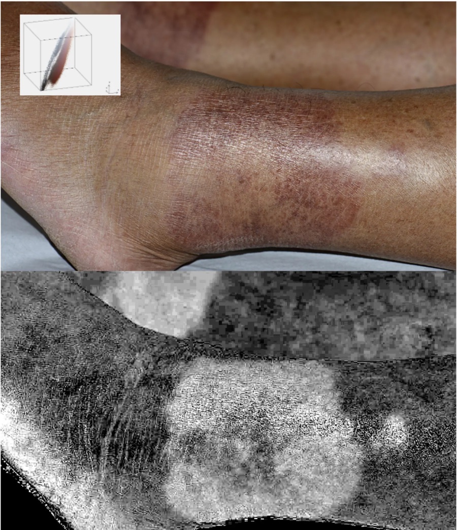
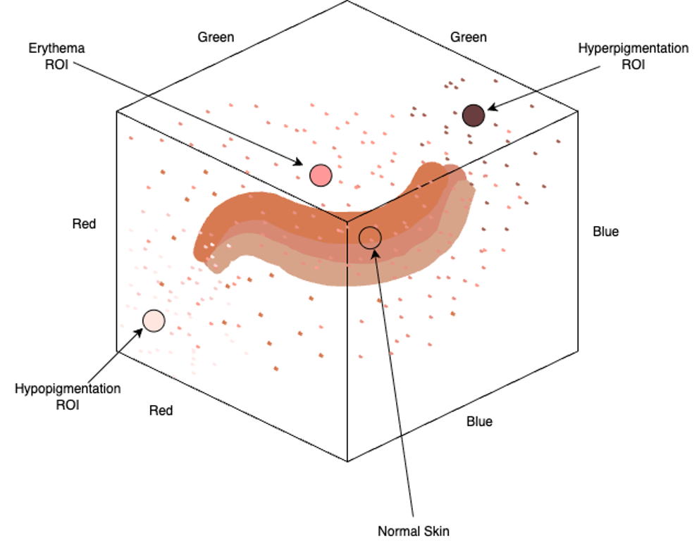
Wide-field and microscopic multispectral, thermal, and color imaging of skin, surfacing vasculature, pigment, and inflammation in ways the human eye cannot.
Devices
Custom and off-the-shelf capture hardware, from radiometric thermal cameras to multispectral rigs, adapted for reproducible skin measurement.
Engineering
Applying mechanical and optical engineering principles to solve problems in the skin, designing novel devices, instruments, and imaging systems.
Clinical Applications
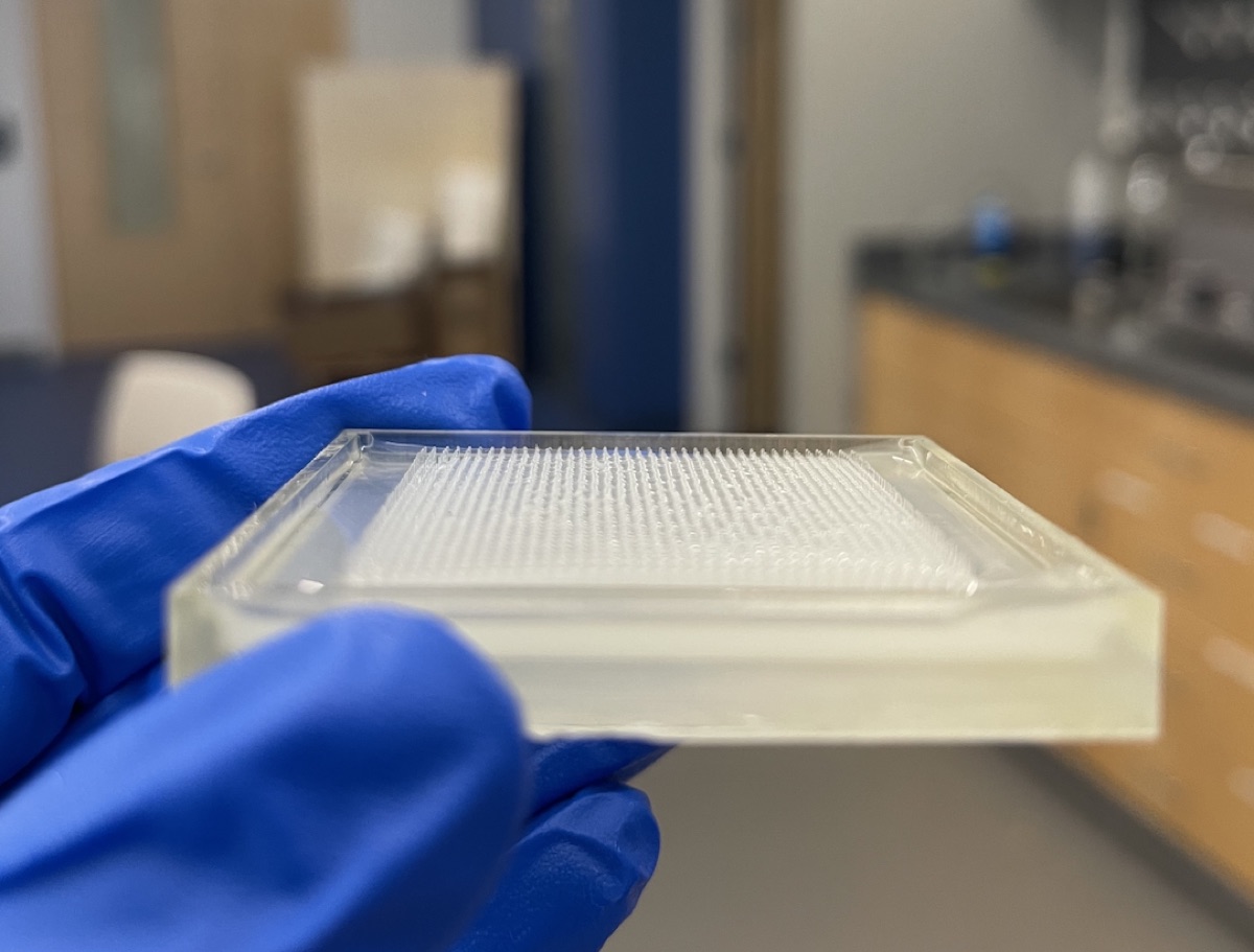
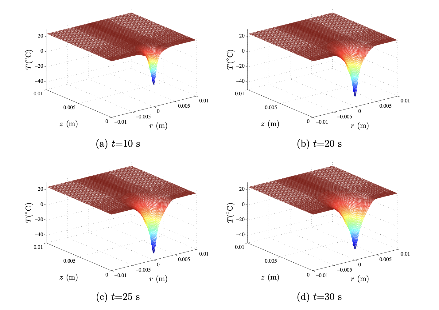
Translating imaging and devices into care, quantifying erythema, guiding diagnosis, and developing low-cost treatments such as fractional cryotherapy for field cancerization.
Active Areas of Research
Our team
The researchers and collaborators behind the IDEAS Lab.
Principal Investigator
William Lewis, MD
Principal Investigator · Beth Israel Deaconess Medical Center
William Lewis is an Instructor in Dermatology at Harvard Medical School and Beth Israel Deaconess Medical Center, where he directs the IDEAS Lab. His work bridges biomedical optics, mechanical and device engineering, and clinical dermatology, with a focus on low-cost, accessible tools for both imaging and treating skin.
He earned his BA summa cum laude from Boston University and his MD from Harvard Medical School, where his research at the Wellman Center for Photomedicine spanned photodynamic and laser therapy and the optical imaging of skin and vascular tissue. He completed internal medicine training at UCLA and dermatology residency at the University of Pennsylvania, both in global-health pathways that included clinical work in Malawi and Guatemala. Since returning to Boston in 2024, he has built a program in smartphone-based imaging of erythema in skin of color, micro-fractional cryotherapy, and new approaches to clinical skin microscopy. His work in this area has been recognized by the Skin of Color Society. He also serves as Director of Medical Student Research in Dermatology at BIDMC.
Current Team
Daniel Cubillos Rojas-Alejandro, MD
Research Fellow · 2026–2027
Imaging & image processing · Bogotá, Colombia (prev. Purdue)
Janelle Clovie
Medical Student
Boston University School of Medicine
Amr Seifelnasr
Researcher · UMass Lowell
Fractional cryotherapy modeling
Majd Elhachem
Researcher
Device design & testing · fractional cryotherapy
Collaborators
Walfre Franco, PhD
Collaborator
Chair, Biomedical Engineering · Francis College of Engineering, UMass Lowell
Alumni
Dalton Driscoll
Alumnus
Now: PhD student, Biomedical Engineering · UMass Lowell
Sophie Numan
Alumna
Research Assistant
Luxanna Sands
Alumna
Now: PhD student, Biomedical Engineering · University of Maryland
What's happening
Recent recognition, presentations, and milestones from the lab.
-
SPIE Photonics West 2026
Congratulations to Dalton Driscoll on acceptance to present "Smartphone Skin Erythema Imaging" at SPIE Photonics West 2026!
-
BMES 2025
Congratulations to Luxanna Sands on presenting "Development of a Fractional Cryotherapy Device for Actinic Field Cancerization" at BMES 2025!
-
Best Presentation · 2025
Congratulations to Dalton Driscoll on winning Best Presentation at the Francis College of Engineering 2025 Student Research and Community Engagement Symposium!
Selected work
Selected publications from the lab and its collaborators.
-
Smartphone Imaging of Subcutaneous Veins
-
RGB Skin Erythema Imaging
-
Tissue Autofluorescence is Correlated with Intima and Media Thickness in Atherosclerotic Human Aorta
Work with us
Interested in collaboration, joining the lab, or learning more?
Email: wlewis2@bidmc.harvard.edu
Location: Department of Dermatology, Beth Israel Deaconess Medical Center · Boston, MA